0
0Anatomy:

Fig. 1. Respiratory system
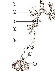
Fig. 2. The lower respiratory tract, or "Respiratory Tree"
- Trachea
- Mainstem bronchus
- Lobar bronchus
- Segmental bronchus
- Bronchiole
- Alveolar duct
- Alveolus
In humans and other mammals, the anatomy of a typical respiratory system is the respiratory tract. The tract is divided into an upper and a lower respiratory tract. The upper tract includes the nose, nasal cavities, sinuses, pharynx and the part of the larynx above the vocal folds. The lower tract (Fig. 2.) includes the lower part of the larynx, the trachea, bronchi, bronchioles and the alveoli.
The branching airways of the lower tract are often described as the respiratory tree or tracheobronchial tree (Fig. 2). The intervals between successive branch points along the various branches of "tree" are often referred to as branching "generations", of which there are, in the adult human about 23. The earlier generations (approximately generations 0–16), consisting of the trachea and the bronchi, as well as the larger bronchioles which simply act as air conduits, bringing air to the respiratory bronchioles, alveolar ducts and alveoli (approximately generations 17–23), where gas exchange takes place. Bronchioles are defined as the small airways lacking and cartilaginous support.
The first bronchi to branch from the trachea are the right and left main bronchi. Second, only in diameter to the trachea (1.8 cm), these bronchi (1 -1.4 cm in diameter) enter the lungs at each hilum, where they branch into narrower secondary bronchi known as lobar bronchi, and these branch into narrower tertiary bronchi known as segmental bronchi. Further divisions of the segmental bronchi (1 to 6 mm in diameter)[7] are known as the 4th order, 5th order, and 6th order segmental bronchi, or grouped together as subsegmental bronchi.
Compared to the, on average, 23 number of branchings of the respiratory tree in the adult human, the mouse has only about 13 such branchings.
The alveoli are the dead end terminals of the "tree", meaning that any air that enters them has to exit via the same route. A system such as this creates dead space, a volume of air (about 150 ml in the adult human) that fills the airways after exhalation and is breathed back into the alveoli before the environmental air reaches them. At the end of inhalation, the airways are filled with environmental air, which is exhaled without coming in contact with the gas exchanger.
Ventilatory volume:
The lungs expand and contract during the breathing cycle, drawing air in and out of the lungs. The volume of air moved in or out of the lungs under normal resting circumstances (the resting tidal volume of about 500 ml), and volumes moved during maximally forced inhalation and maximally forced exhalation are measured in humans by spirometry. A typical adult human spirogram with the names given to the various excursions in volume the lungs can undergo is illustrated below (Fig. 3):

Fig. 3 Output of a 'spirometer'. Upward movement of the graph, read from the left, indicates the intake of air; downward movements represent exhalation.